Photocopying Your Bones in 3D: Making 3D Printing for Surgical Planning A Reality in Trinidad and Tobago
Nov 26, 2019 | Views:219136 |
 Print Version
The use of 3D-printed anatomical models to visualise complex pathology is becoming more commonplace in orthopaedic surgical practice. 3D-printed models are created from high resolution patient scan data (such as CT and MRI) and can be used for preoperative planning, collaboration within the surgical team and as an aid to communication with the patient during consultation. 3D anatomical models can also be used for surgical education allowing for training on physical models of fractures, tumours and other abnormalities.
An additional advantage of patient specific 3D-printed anatomical models is the cost-savings associated with reduced surgical time. The use of 3D printed anatomical models results in better and shorter surgeries at lower costs. Some studies have shown that surgical time can be reduced by 30-60 minutes on average, up to a maximum of 5 hours depending on the procedural complexity. The ability to create 3D-printed anatomical models is now available to surgeons in Trinidad and Tobago via the Design and Innovation Group led by Dr. Umesh Persad, Associate Professor, Design and Manufacturing Systems at The University of Trinidad and Tobago (UTT).
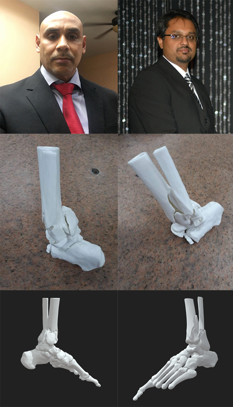
In close collaboration with Dr. Marlon Mencia, Lecturer in Trauma and Orthopaedics, The University of the West Indies (UWI) and Consultant Orthopaedic Surgeon at Westshore Medical Private Hospital, a 3D-printed model of a patient’s pilon fracture was produced for an initial research case study. A pilon fracture is a break that occurs at the bottom of the tibia (shinbone) as the result of a high-energy injury. The bones are crushed or split into several pieces due to the high-energy impact and surgery is required to restore anatomical alignment.
The patient’s high-resolution CT image data was used to reconstruct a 3D model that was printed at Qualitech Machining Services Limited, a key collaborator of the Design and Innovation Group. The model was used for preoperative planning which allowed for better spatial appreciation of the fracture pattern and helped with planning the surgical approach. The surgical team felt that there was significant benefit in having a common tangible reference model of the patient’s anatomy. The model also facilitated the preoperative selection of the implants required for surgical stabilisation, thereby reducing both operating time and radiation exposure during surgery. The model was particularly useful in explaining to the patient the nature of the injury and what would be done at surgery, enabling informed consent.
Dr. Mencia is hopeful that the benefits of 3D-printed anatomical models will lead to improved clinical outcomes, and 3D preoperative models are currently being used for other cases. Dr. Persad is also hopeful that making such technology available to the local and Caribbean populations would directly impact the quality and cost effectiveness of medical care and surgical intervention. It also speaks to the impact that UTT is having in fulfilling its mission to improve the quality of life in Trinidad and Tobago via research and entrepreneurship.
More Results
