
UTT Design and Manufacturing Engineering researchers develop new medical imaging algorithm for brain tumors
Nov 14, 2017 | Views:211893 |
 Print Version
Amid the variations of the cancer disease, brain tumors account for the majority of deaths among young people. To diagnose and treat this deadly disease effectively, analysis of hundreds of medical images such as Magnetic Resonance Imaging (MRI) scans is usually performed. However, the analyses of these scans are still mainly performed manually, making the procedure not only very tedious and time-consuming for doctors, but also error prone and non-repeatable. Attempts have been made to automate this procedure by performing image processing techniques such as thresholding, region-growing, unsupervised learning like k-means or fuzzy c-means clustering, and supervised learning e.g. support vector machines. Some require human interaction. The techniques may be applied on one or more MRI sequence scans. Unfortunately, these automated attempts still result in a high level of error, and more computationally complex algorithms do not guarantee an increase in accuracy.
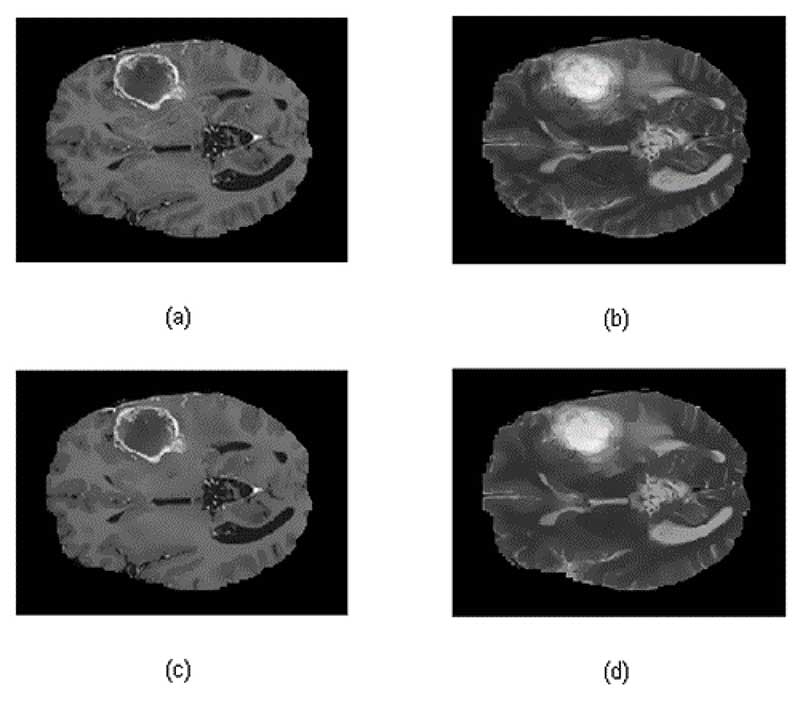
Amrit Hanuman (Research Assistant), Dr. Umesh Persad (Assistant Professor and Principal Investigator) and Dr. Ken Sooknanan (Assistant Professor ICT and Co-Investigator) developed a novel, fully automatic brain tumor segmentation and volume estimation method using simple techniques on T1-contrasted and T2 MRIs. This new approach implemented five main steps: preprocessing using anisotropic diffusion, segmentation of tumor regions using k-means clustering, region combination using logical and morphological operations, error checking using temporal smoothing, and volumetric measurement. When compared with five state-of-the-art algorithms, the proposed algorithm outperformed those in past works. Advances were seen by its noise reduction, increase in accuracy and closeness to actual tumor volume.
Further work would involve reduction in the execution time and catering for data that is not skull-stripped or aligned. Additionally, an alternate, semi-automatic option can be developed to allow doctors to draw a region of interest for segmentation on the MRI. Finally, the ultimate goal is to implement the algorithm in radiological analysis software for clinical use to directly impact the lives of patients.
More Results


